

Romexis is the world's leading all-in-one software for dental practices and clinics: Romexis links all compatible imaging devices and networks them with the practice software.
This not only makes diagnostic processes easier and more convenient but also offers smart options for optimising and digitising entire workflows.
- All imaging units are combined into one comprehensive software platform
- A future-proof concept – a solution with excellent upgradeability
- Reliability through proven technologies
- Suitable for all sizes of practice, even those with several locations, up to large clinics
- Integrated document management
- Central, device-independent diagnostic image archive
- Extensive tool palettes for viewing, editing, measuring and evaluating
- Overlay option for DVT recordings and support for diagnostic analysis and control
- Software solution that is also open to third-party providers (TWAIN standard)
- Compatible with macOS* and Windows

Romexis 7 – The New Generation, Powered by Integrated AI.
With powerful AI capabilities built in as standard, Romexis 7 takes care of time-consuming routine tasks, reduces manual work and streamlines your workflow. So you can spend less time on routine tasks and more time focusing on what truly matters: delivering outstanding care to your patients.
The new AI functions in detail

Romexis® software uses AI-powered tools to automatically segment CBCT images, significantly reducing the time required compared with manual segmentation.
Key anatomical structures such as teeth, nerves, jaws, airways, and paranasal sinuses are automatically identified and visualized. This simplifies diagnostic analysis and treatment planning while also enhancing patient consultation through informative 3D visualizations.
The segmented objects can also be exported as STL files and used directly in digital workflows and for 3D printing.
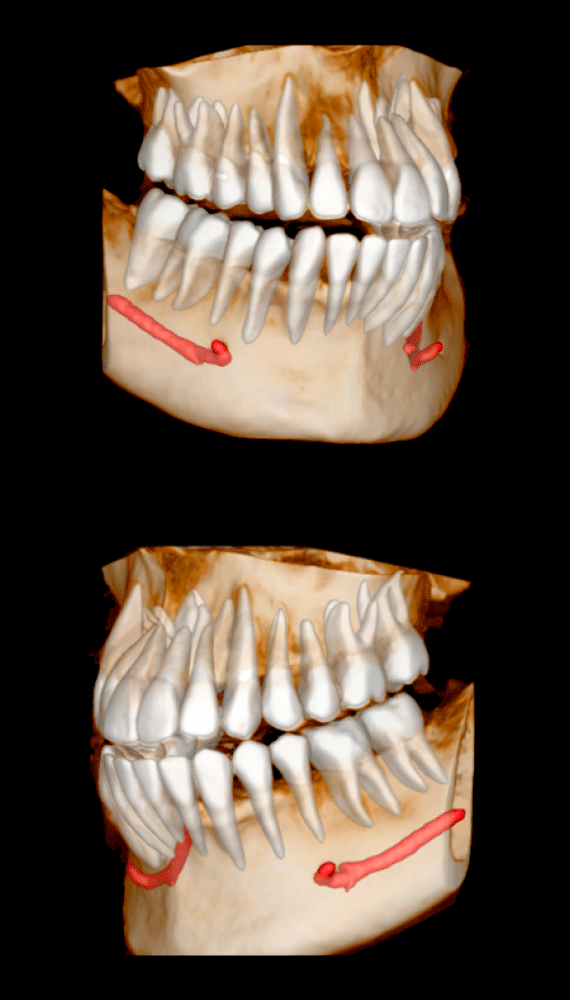
Romexis intelligent technology automatically identifies the mandibular canal in CBCT images and clearly visualizes its course.
This is particularly important for procedures such as implant surgery or wisdom tooth extraction: the nerve canal is clearly displayed in the CBCT images, enabling precise treatment planning.
If necessary, the course of the nerve can be adjusted manually.
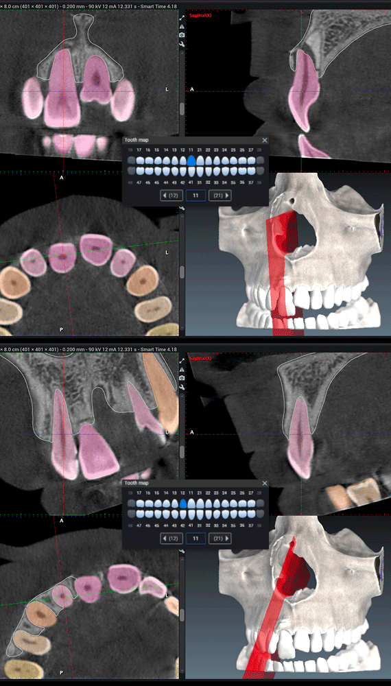
Romexis automatic anatomy recognition makes navigating 3D images particularly convenient. Simply select a tooth, and the software automatically centers all views on the selected tooth.
The selected tooth remains centered even during a full 360-degree rotation, allowing you to obtain the best possible viewing angle for diagnosis and treatment planning at any time.
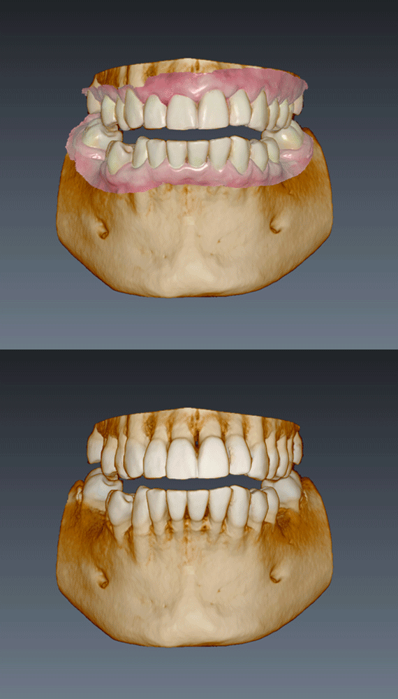
Romexis automatically combines CBCT data and intraoral scans into a single digital model.
This saves time when creating virtual setups for implant surgical guide design or orthognathic surgery planning.
Romexis also enables the creation of a highly detailed segmented tooth model by merging the crown from an intraoral scan with the root structures from CBCT images.
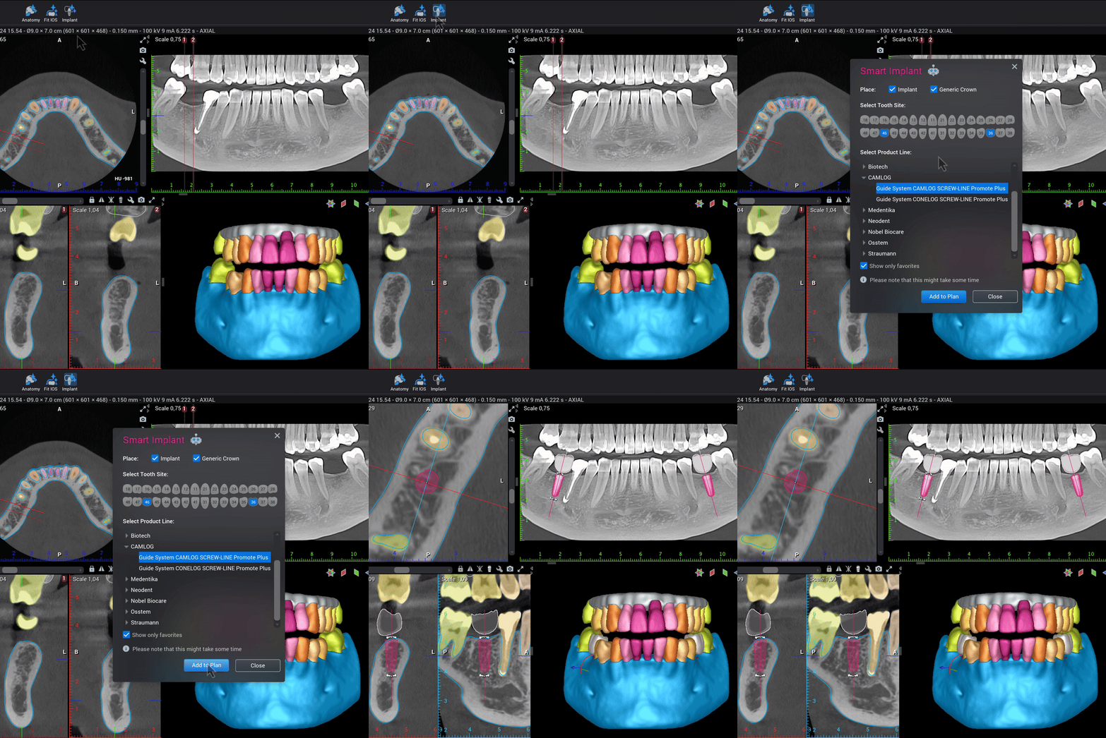
With the Romexis® implant planning module, you can create implant plans particularly efficiently. Simply select your preferred product line from our implant library, which includes products from more than 130 manufacturers.
The software automatically suggests a suitable implant position, creates a virtual crown, and takes anatomical structures such as bone, adjacent teeth, and the mandibular nerve into account.
You can then review the proposed plan and adjust it individually as needed.

With the Romexis® 3D Implant Guide module, you can design surgical guides in just a few steps.
Thanks to an integrated library containing more than 50 implant guide kits from various manufacturers, the software automatically detects the guide area and generates a suitable guide in just a few steps.
The guide can then be customized with support bars, inspection windows, or individual labeling. Teeth that have already been extracted can also be removed from the treatment plan with a single click, without requiring a new scan.

Romexis uses advanced AI technology to automatically identify and analyze anatomical landmarks in 3D images.
This enables analyses to be generated more quickly while reducing manual work to a minimum.
At the same time, users retain full control, as all results can be reviewed and corrected if necessary.

Romexis uses artificial intelligence to automatically identify teeth in intraoral X-ray images and assign them to the correct tooth numbers. This significantly reduces manual work and saves valuable time.
In addition, the software automatically rotates each image so that the teeth are always displayed in the correct orientation.
When study templates are used, the images are automatically placed in their designated positions. (Romexis 6.5.2.)

The AI-powered assistant provides precise and concise answers to questions about using the software and guides you through step-by-step instructions whenever you need assistance.
The simple top-down implant workflow.


Acquire a CBCT scan.


Automatic matching of the intraoral scan and CBCT scan.


Select the desired implant and the appropriate sleeve from the Romexis library. Romexis automatically suggests the optimal implant position while taking relevant anatomical structures into account.


Design the implant surgical guide directly in Romexis software. The software automatically generates an open STL file for the design and enables the creation of additional tooth-supported and mucosa-supported guides.

Print the completed surgical guide using a 3D printer.
Software with a multitude of possibilities

Romexis is the all-in-one software for any size of practice and all dental specialties.
Perfectly matched to the entire KaVo ProXam imaging portfolio, both small practices and clinics benefit from Romexis. The all-in-one software is impressive in its versatility and is based on a concept that means you have a full range of functionality both now and in the future: thanks to its compatibility with Windows and macOS as well as the open interfaces – also compatibility with third-party solutions – Romexis is a flexible option that can be adapted quickly and easily to suit your individual requirements.
Anyone looking for versatile and intelligent software with uncompromising quality and reliable premium features in the field of diagnostics will love Romexis, especially in combination with the new ProXam diagnostics portfolio, for long-lasting, sustainable benefits.

DVT image

2D panoramic image

3D implant
All practices and clinics
- Private practices with one treatment room all the way up to large, multi-surgery practices
- Practices with multiple locations
- Medium-sized clinics
- Hospitals and universities
All specialities
- Radiology
- Implantology
- Dental prosthetics
- Orthodontics
- Endodontics
- Periodontology
- Aesthetic dentistry
All data
- DVT imaging
- 2D X-ray images
- Photos and videos
- Intraoral 3D scanners
- TWAIN units
All platforms
- Native support for Windows and Mac (Some features are only supported on Windows operating systems.)
Users
worldwide trust in Romexis.
Years
successfully used in practices and clinics.
Images
of more than 1.4 Million patients processed per year, making up the largest Romexis-linked network within a practice.
The universal software platform for the dental imaging process.
Romexis supports dentists in the following steps:
- Image creation
- Diagnostic evaluation
- Editing and storage of 2D and 3D X-ray images and intraoral scans
- Processing CAD/CAM data
Romexis specifications & system recommendations
Romexis offers a variety of reliable tools that support either detailed treatment planning or the design of drilling templates, splints, and tooth models, as well as restorations for in-house 3D printing or milling.
With its large number of integrated image processing programmes and easy integration to practice management systems – also from third parties – Romexis is an open, comprehensive, and future-proof software solution. All 2D, 3D and CAD/CAM data can be exported or imported in standard format at no additional cost. Romexis supports the DICOM (Digital Imaging and Communications in Medicine) standard, which can also be connected to hospital systems.
With Romexis, it is easy to create professional, high-quality diagnostic documents or radiology reports and send them to referring doctors. Multi-page documents of any format can also be attached directly to patient files. This is not only quick and easy, but also facilitates the storage of Ceph tracings, bank transfers and other important documents.
With Romexis, it is easy to create professional, high-quality diagnostic documents or radiology reports and send them to referring doctors. Multi-page documents of any format can also be attached directly to patient files. This is not only quick and easy, but also makes it easier to store Ceph tracings, bank transfers and other important documents.
From surgical planning to template-supported surgery: the Romexis 3D Implant Planning module provides all the necessary tools and technologies for digital implantology. By visually laying the crown and tooth model over the DVT data, users can create a complete, virtual setup from a prosthetic and surgical point of view for optimal positioning of the implant.
The software features an intuitive user interface and optimized standard workflows, allowing daily tasks to be completed quickly and with as few clicks as possible. AI-powered tools automate repetitive tasks and minimize manual effort, further streamlining workflows.
To support users, Romexis also provides an extensive library of instructional videos as well as a large language model-powered Smart Chat, making it easier to use the software quickly and efficiently.
As cyber threats are increasing in volume and complexity, the importance of data privacy and security cannot be overstated. Keeping personal and patient data safe should be a top priority for all dental organisations and considered in all aspects of their operations. We have built security into our Romexis software since day one – making it easy for all users to follow responsible practices and meet tightening regulatory requirements.
Powerful features for precise diagnoses in all specialist areas.
Powerful optimisation and analysis tools form the foundation for precise diagnoses in all specialist areas. In order to ensure maximum flexibility, Romexis naturally adapts to personal needs and individual practice or clinic requirements when it comes to imaging.
As Romexis is modular software, you can start with the features you need and add further optional software modules later. You can expand from 2D to 3D imaging and subsequently add CAD/CAM capabilities, complete implant workflows, or virtual planning for orthognathic surgery. The software is regularly updated with new features and improvements.
- Extensive selection of reliable tools for viewing, editing, measuring, drawing and evaluating recordings
- The Romexis 3D rendering function provides a perfect overview of the anatomical situation: 3D images can be viewed from different perspectives, and converted into panoramic images or even cross-sectional layers – for a comfortable and patient-friendly 3D diagnosis
- Superimposed DVT recordings offer users a diagnostic tool for analysis, control, and before/after comparisons
Overview Romexis Modules
Romexis 2D Standard
- 2D image acquisition with imaging devices
- TWAIN acquisition with 3rd party imaging devices
- Support for intraoral, panoramic, and cephalometric X-ray images, as well as 3D snapshots and photos
- Image processing, measurement and annotation tools
- Support for image study templates
- AI-based automatic tooth number detection for intraoral x-rays
- Automatic rotation of intraoral X-rays based on AI-detected tooth numbers
- Automatic placement of intraoral X-rays in the study template based on AI-detected tooth numbers
- Customisable prefilters for all image types
- Multi-page printing with customer branding
- Imports and exports: DICOM, JPEG, PNG, TIFF, BMP
- Exports with free Romexis Viewer
- Video, PDF, and document attachments
- DICOM Media Storage (DICOMDIR)
- User management and permissions, including audit trails
- Finding patients by image type, date, or comment
- Assigning patients to users
- Launch for external applications
Romexis Smile Design
- Photorealistic simulation of new smiles
- Teeth silhouette with teeth shape library, creating custom shapes
- Grid for edentulous cases
- Tooth shade guide and selection
- Facial analysis tools
- Mapping facial and intraoral photos
- Exports to CAD/CAM or other 3D systems
- Automatic smile design report and custom printing
- Includes Romexis 2D Standard
Romexis 3D Standard
- Image acquisition with CBCT units
- MPR views (axial, sagittal, coronal)
- 3D rendering views
- Automatic panoramic views
- Cross-sectional views
- Automatic segmentation of teeth, maxilla, mandible, airways and sinuses
- Automatic tooth number recognition
- Automatic nerve canal tracing
- Automatic CBCT and IO-scan mapping
- Tooth number-based navigation
- Pseudopanoramic and cross-sectional views
- Image processing, annotation, and measurement tools
- Imports: DICOM, STL
- Exports: DICOM, STL, OBJ, PLY
- Converting CBCT images to STL files
- Segmentation of jaws and tooth
- Segmentation of airways
- Segmentation using region growing
- Nerve canal tracing and root canal marking
- Mapping CBCT images and dental models or any STL file
- Creating virtual cephalometric images
- TMJ views
- Superimposing two CBCT volumes
- Creating 2D snapshots and 2D slice stacks
- Support for Planmeca 3D photos
- Mapping CBCT images with 3D or 2D photos
- Comparison of two 3D photos or surface scans
- Distance and angle measurements between two surfaces
- Shaping 3D photos
- Two surface measurements
- Launch for external applications
- Includes Romexis 2D Standard
Romexis 3D Implant
- Automated implant plan and virtual crown proposals
- Implant planning tools (alignment, implant extension, implant safety areas)
- Implant centric views
- Implant libraries featuring +130 manufacturers
- Abutment libraries and a generic abutment designer
- Generic crown library
- Implant verification tool
- Automatic implant reports
- Includes Romexis 2D Standard
- Includes Romexis 2D Implant
- Includes Romexis 3D Standard
Romexis 3D Implant Guide
- Implant guide design tools for tooth supported guides
- Implant guide design tools for mucosa supported guides
- Automatic guide area detection
- Automatic tooth extraction
- Presets for 3D printers
- Automatic Implant and sleeve reports
- STL export for guides
- Includes Romexis 2D Standard
- Includes Romexis 3D Standard
- Includes Romexis 3D Implant
Romexis CMF Surgery
- Workflow Wizard
- Automatic segmentation of teeth, maxilla, mandible, airways and sinuses
- Automatic landmark detection
- Automatic CBCT and intraoral scan mapping
- Placing and defining anatomical landmarks
- Dynamic measurements and analyses
- Head orientation tool for manual adjustment
- Viewing bone segment projections in slice views
- Planning maxilla osteotomies: Le Fort I, One-piece, Two-pieces and Three-pieces
- Planning mandible osteotomies: BSSO Hunsuck and Obwegeser, Inverted L, vertical ramus and Genioplasty
- Showing osteotomy lines dynamically in slice views
- Showing marked nerves during osteotomy planning
- Showing segmented teeth
- Fitting the target model
- Movement planning with presets for the most used movement types
- Preoperative to virtual plan superimposition
- Creating intermediate and final splints, open STL export
- Export of postoperative models in open STL format
- Extensive surgery reports
- Includes Romexis 2D Standard
- Includes Romexis 3D Standard
Romexis 3D Cephalometry
- Placing anatomical landmarks in 3D view or on 2D slice views
- Head orientation tool for manual adjustment
- Dynamic measurements and analyses
- Measurement table for comparisons against the norms
- Landmark, plane, and measurement visualisation in 2D views and 3D
- 3D photo visualisation
- Analysis types: TFA Perrotti analysis, Orthognathic Surgery analysis
- Includes Romexis 2D Standard
- Includes Romexis 3D Standard
Romexis VR
- Launch and view 3D patient data in virtual reality
- Examine patient anatomy in true three dimensions
- Plan implant treatments using extensive implant libraries
- Engage patients in immersive experience
Romexis CAD/CAM
Main features
- Scanning with Intraoral scanner KaVo ProXam iOS**
- Presets for most common workflows
- Scanning different bites
- Buccalless alignment for full arch cases - Taking 2D snapshots with the scanner
- Model editing, orientation and viewing
- Shade assistant - Automatic AI-based analyses and measurements
- Tooth width and arch length measurements
- Bolton and Space analyses
- Overjet and overbite measurements - Analysing dental models
- Contact map and undercut calculations
- Tooth width, arch length, and free measurements
- Overjet and overbite measurements
- Bolton and space analyses
- Comparison of scans - Model base creation (solid and hollow)
- Marking the margin lines for multiple teeth
- Imports and exports: STL, PLY
- Export: PlanCAD Premium, 3Shape and exocad formats
- Send: DDX Cloud, HeySmile and SIMtoCARE
- Creating lab order forms (PDF)
- Includes Romexis 2D Standard
**Support for the Windows operation system only
Romexis Ortho Simulator
- Automatic one-click simulation of the teeth position after the aligner treatment
- Automatic orientation of the scans
- Automatic segmentation of crowns from the intraoral scan
- Tools for modifying the automatic segmentation and labelling
- Automatic detection of the tooth axis
- Tools for adjusting the tooth axis, both mesial-distal and long axis
- Manual movement and rotation of the tooth
- Possibility to reset the segmentation and the simulation
- Taking 2D snapshots
- Includes Romexis 2D Standard
- Includes Romexis CAD/CAM Scan
DICOM Print
- DICOM-Print SCU
DICOM Full
- DICOM-Print SCU
- DICOM-Storage SCU
- DICOM-Worklist SCU
- DICOM Query/Retrieve SCU
- DICOM-Storage Commitment
- DICOM MPPS
DICOM Dental PACS
- DICOM Storage SCP
- DICOM Query/Retrieve SCP
- Access control
- Event logging
- Resend capability
- Includes DICOM Full
Romexis Specifications & System requirements
The implant library – the best conditions for top-down implant planning.
The Romexis implant library includes numerous realistic implant models and sleeves for drilling templates. In addition, the software grants access to implant models from over 100 manufacturers. After completing the implant planning, Romexis can produce the desired drilling template with just a few clicks.
Romexis Viewer
Full-featured application for viewing Romexis 2D and 3D images. The application can be exported and sent together with images from the Romexis software.


