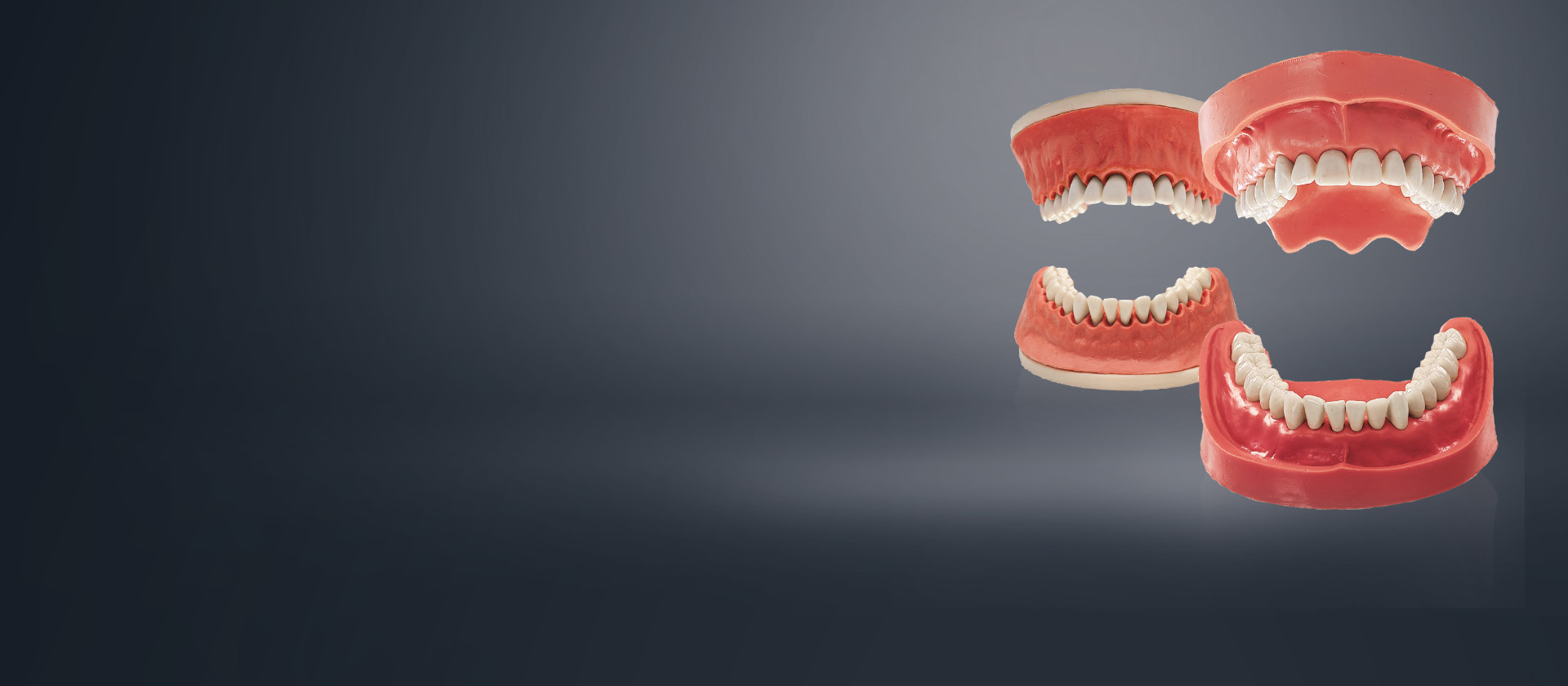
Learning to treat periodontal disease with models as realistic as the real thing.
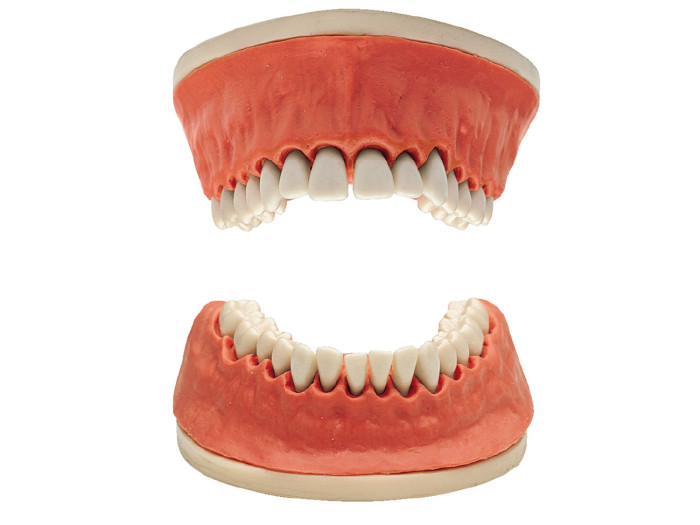
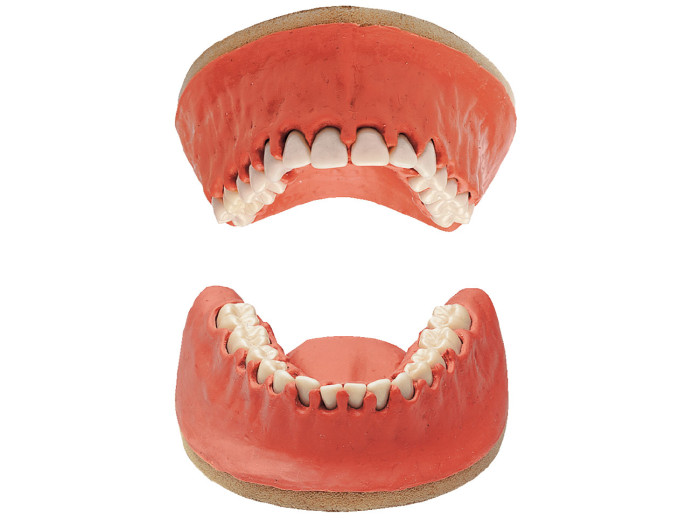
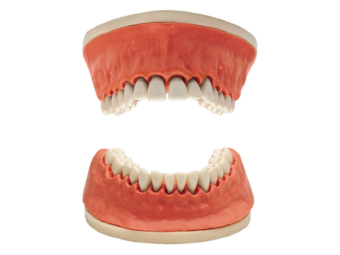
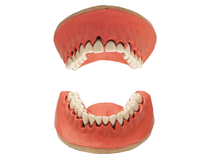
KaVo tooth models for periodontics have particularly soft gingiva and are equipped with root teeth so that students can carry out periodontal treatment exercises as realistically as possible during their course. The KaVo root teeth make scaling, root planning and GTR exercises as realistic as possible.
- Tooth models for realistic periodontal diagnostics and therapy
- High-quality, precision KaVo workmanship
- Convincingly realistic tooth models for periodontics
- Comes with soft gingiva and root teeth
Equipment
- Delivery mounted in labelled, reusable individual transparent boxes.
- Root teeth made of melamine resin with natural root.
- PVC gingiva in natural colour or transparent.
- Baseplate for fixing model in patient simulator (using M6 screw or magnet).
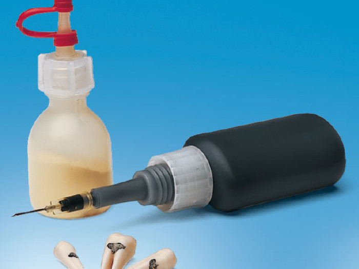
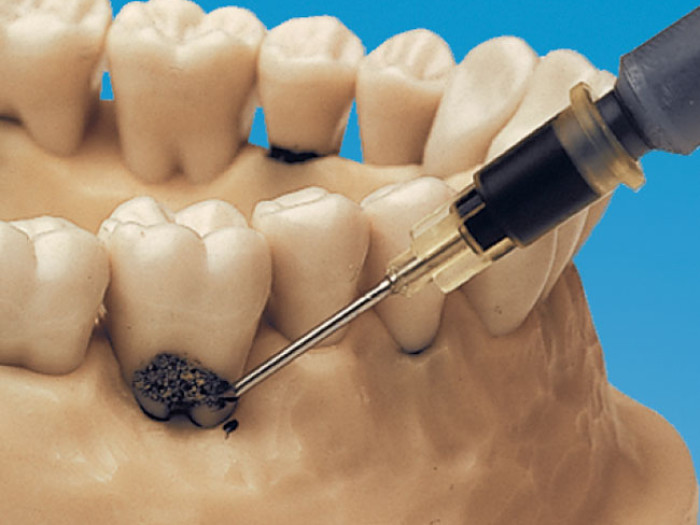
- Optional set of artificial tartar.
Benefits
- Preclinical training of periodontics treatments under realistic conditions.
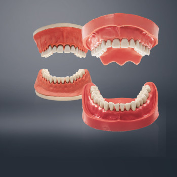
- Training on 4 different, advanced periodontal states for adults.
- Scaling training using the tartar set.
Periodontics study and tooth models
The KaVo periodontics models were developed in cooperation with the Foundation for Clinical Research of the University of Bern and are available in four different status types. The following work can be practised and documented with the aid of the KaVo periodontal set type forms:
- Determination of the periodontal status
- Preparation of a treatment plan
- Scaling / Curettage
- Root planing
- Root amputation
For early exercises, transparent gingivae are also available. Further possible exercises can be performed using the KaVo tartar set.
KaVo root teeth offer the possibility of Scaling, Root planing and GTR exercises.
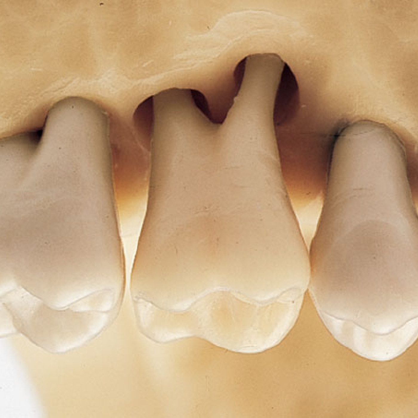
Localised periodontal disease with intra-osseous components can be diagnosed.
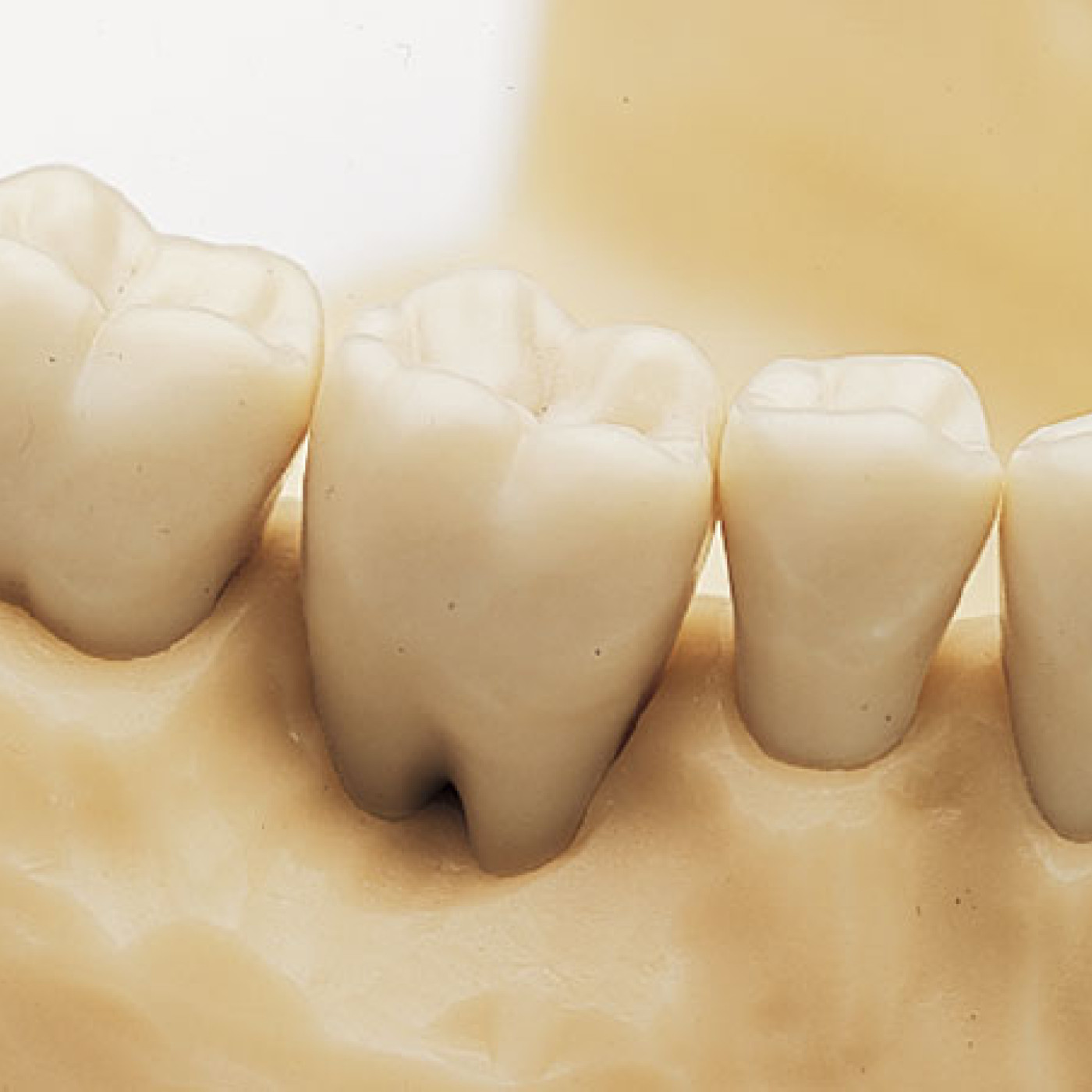
Clearly open furcations (Class II) are to be found in the mandibular.
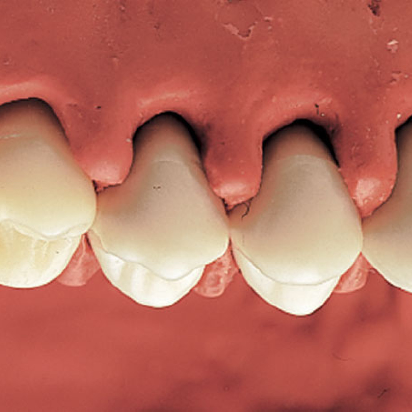
Hyperplasia primarily in the interdental spaces indicated.
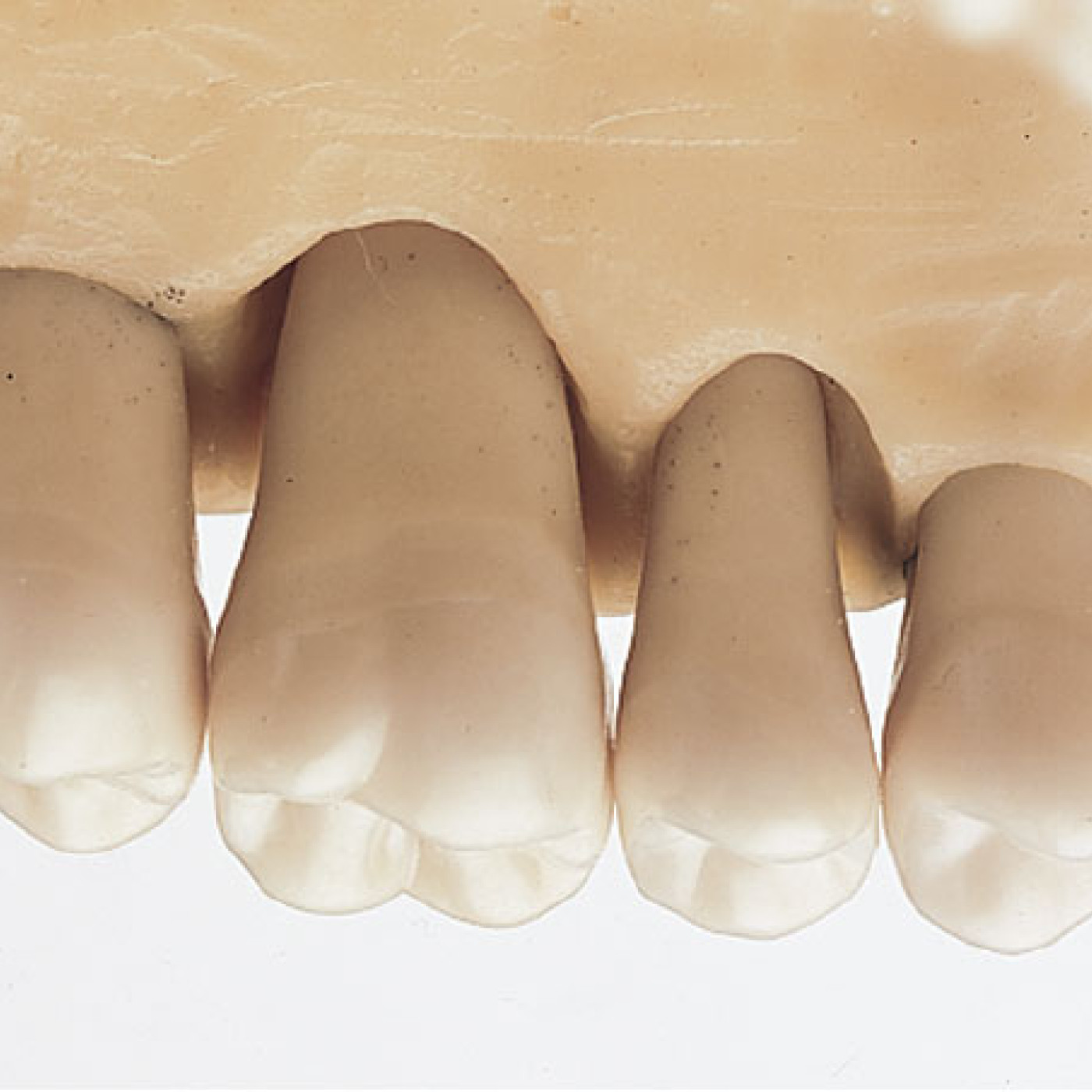
Periodontitis well-advanced in the molar region.
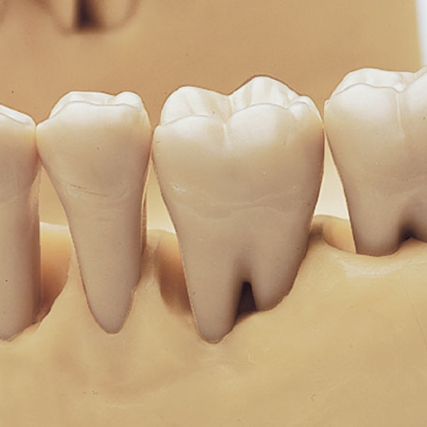
Universal early furcation periodontitis on teeth 36 and 37.
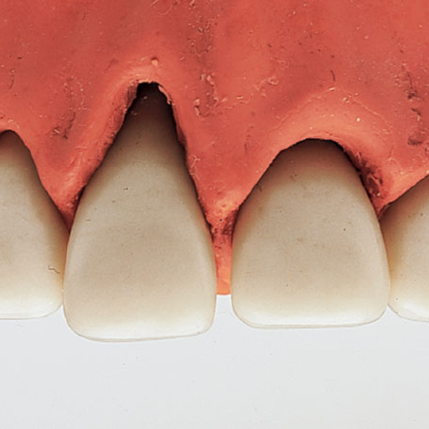
„Cleft-like“ recessions visible in the upper as well as in the lower jaw.
Helpful supplements
Holding screw aids for periodontal study models & simulated calculus kit.
